

Digital Photographic Monitoring
 Melanoma is a skin cancer that is known to change over time, often slowly in its initial stages. In contrast, very few normal moles change over a short period of time. This allows doctors to use the technique of short term digital photographic monitoring to aid in the diagnosis of early melanomas.
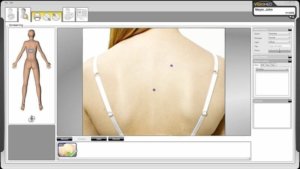
Digital photographic monitoring of a skin lesion may be recommended by your doctor for lesions that are only marginally atypical and do not fulfil the criteria for performing a biopsy. In this technique, a skin lesion which may appear as a slightly atypical mole on dermoscopic examination is photographed and the image stored on a computer. The patient is then asked to return should a change be noticed, as well as after 3 months, to repeat the photograph. The two images are then compared and if there is a slight change in the lesion then it is excised for biopsy and microscopic analysis.
The use of this technique has been shown to improve the diagnostic accuracy for the detection of early melanomas. If your doctor has recommended this method of monitoring it is essential that you remember to return for your 3 monthly follow-up appointment.
Illustrated below is a pigmented lesion at the time of initial examination with clinical and dermoscopic photos, and the same lesion 3 months later. There is a change in the follow-up dermoscopic image. The lesion was excised to reveal an early (in-situ) melanoma. It is difficult to find clear clues to melanoma in either of the images, however, the change observed has allowed the diagnosis to be made.
Your doctor will be able to diagnose most skin cancers during the consultation, however, occasionally a biopsy will be required to check the spot under the microscope in order to make the diagnosis. This is usually able to be completed on the day of your consultation.
Once the area is numbed with local anaesthetic a small sample of skin is removed and sent to a specialist dermatopathologist who can diagnose the skin spot under the microscope.
Sometimes a larger piece of skin needs to be removed for the biopsy and will require stitches – this is particularly the case for moles that are suspected of being melanoma.
Results are available one week later and an appointment arranged if further treatment is required. Patients are asked to contact the centre initially to discuss their result over the telephone. You are welcome to keep a copy of your report, which can also be made available by your GP.